Il popolo dei runnersè sempre più popoloso e con esso anche coloro i quali soffrono di patologie correlate come la periostite tibiale.
Che cosa sia realmente questa patologia e come differenziarla da altre cause di dolore tibiale è ancora oggi una sfida…
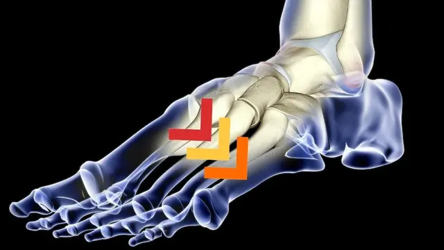
In realtà il termine “periostite tibiale”, preso così com’è, definirebbe una patologia infiammatoriaa carico del periostio, cioè la membranache riveste le ossa. In realtà l’eziopatoenesi della patologia, ciò la causa, non è ancora stata completamente chiarita.
Nel 1986 era già stata proposta una classificazione (fig.1) dove diverse patologie quali periostite, fratture da stress, sindrome compartimentale cronica eintrappolamento dell’arteria poplitea si sovrapponevano in un grande calderone di entità che causano un dolore alle gambe sport-correlato.
 classificazione delle sindromi legate al dolore di tibia")
Attualmente il termine anglossasone per definire questa patologia è medial tibial stress syndrome o MTSS. Con questo termine si definiesce come causa principale proprio lo stress a cui è sottoposto l’osso (tibia) durante la marciao la corsa. I progressi nella tomografia computerizzata, nella MOC e DEXA e nella risonanza magnetica hanno consentito di dimostrare sia la presenza di edema osseo che di sollevamento periostale causato dell’essudato osseo sottostante. Infine sono stati evidenziati segni di riassorbimento osseo della porzione posteromediale tibiale. Comunque, l’accuratezza delle diverse tecniche varia notevolmente e può dipendere dai tempi dell’indagine in relazione all’insorgenza del dolore. Solo un numero limitato di studi istologici ha incluso la biopsia ossea e quest’ultimi confermano positivamente i marcatori di stress osseo. Altri studi di imaging dimostrano lesioni ossee associate a una diagnosi clinica di MTSS in siti come la tibia prossimale e la corticale tibiale anterolaterale, che sono incoerenti con le sedi clinicamente significative di dolore.
Questo, come vedete, solleva domande sulla diagnosi differenziale di MTSS e su la natura della patologia stessa che probabilmente, esattamente come mostra la figura n.1, è un concorso di diverse cause.
Aldilà di queste considerazioni, quello che interessa soprattutto ad un runner è il tempo di recupero.
I tempi di recupero tendono ad essere lunghi. In un recente studio i soggetti campione sono tornati a correre per almeno in media 115 giorni. 4 mesi sono tanti per chi ama questo sport, questo dato deve far riflettere su quanto sia implortante una corretta diagnosi ed un piano terapeutico adeguato. Siccome sui trattamenti realmente efficaci c’è ancora molta confusione e poca letteratura solida al riguardo, diversi autori hanno cercato di indagare i fattori di rischio associati alla periostite tibiale (MTSS) al fine di sviluppare screening e contromisure per prevenirla.
Di seguito ti voglio parlare dei principali fattori di rischio, andando ad esaminarli uno ad uno e cercando di farti capire quali di questi siano “affidabili” e quali invece non si sono dimostrati capaci di predirre il possibile sviluppo della patologia.
Fattori di rischio associati allo sviluppo della periostite tibiale (MTSS):
1) navicular drop
Ossia l’altezza dell’osso navicolare (osso del piede primo della fila distale delle ossa del tarso) nella sua posizione neutrale in carico ed in posizione rilassata. Questo test si è dimostrato essere significativamente associato allo sviluppo di periostite tibiale, quindi ad una maggiore differenza di altezza corrisponde un aumentato rischio di MTSS. La soglia critica è circa 10 mm. PROMOSSO!
2) piede pronato
Utilizzando diversi parametri, si può definire un piede in piede pronato, neutrale o supinato. In uno studio riportato da Sharma et al, il tipo di piede è stato classificato in base ai dati della pedana baropodometrica che registrava la quantità di pressione del piede da mediale a laterale durante un’attività a piedi nudi. Questa misura non si è rilevata utile per predirre un rischio di sviluppo della periostite tibiale (MTSS). BOCCIATO!
3) utilizzo di plantari
Sono tre i lavori scientifici che hanno esaminato i runner sull’uso di plantari. Il risultato è che coloro che indossavano ortesi avevano un rischio relativo più elevato di sviluppare periostite tibiale (MTSS). BOCCIATO!
4) indice di massa corporea
In questo caso sonno 5 i lavori scientifici che l’hanno esaminato. Un BMIpiù elevato è significativamente associato allo sviluppo di periostite tibiale (MTSS). PROMOSSO!
5) Dorsiflessione della caviglia (muscolo soleo)
Quattro studi hanno esaminato la relazione tra i gradi di dorsiflessione della caviglia misurata con il ginocchio piegato per diminuire qualsiasi effetto di costrizione del muscolo gastrocnemio. Sono stati testati un totale di 886 persone e non è stato rilevato una correlazione tra questa variabile e lo sviluppo periostite tibiale (MTSS). BOCCIATO!
6) Dorsiflessione della caviglia (muscolo gastrocnemio)
Similmente al muscolo soleo non è stato rilevato una correlazione tra questa variabile e lo sviluppo periostite tibiale (MTSS). BOCCIATO!
7) esperienza
Due lavori scientifici hanno dimostrato su un totale di 182 runner che la possibilità di sviluppare la periostite tibiale (MTSS) è inversamente proporzionale con l’esperienza (espressa in anni) di corsa. PROMOSSO!
8) precedente storia di periostite tibiale
In cinque articoli, su un totale di 515 partecipanti, si è ampiamente dimostrato come chi ha già sofferto di questa patologia sia più esposto a risoffrirne. PROMOSSO!
9) sesso femminile
Il sesso femminile è un significativo fattore di rischio per sviluppare una periostite tibiale (MTSS). PROMOSSO!
10) rotazione esterna dell’anca eccessiva
In questo caso si è calcolato attreverso l’utilizzo del goniometro la massima rotazione esterna dell’anca. Una rotazione esterna eccessiva aumenta la possibilità di sviluppare la periostite tibiale (MTSS). Tale fattore di rischio però è stato dimostrato solo nel sesso maschile. PROMOSSO!
Come hai visto i fattori correlati sono molti e, quindi, è di fondamentale importanza un completo esame fisico per poter individuarli ed impostare uno schema preventivo. I runners che hanno una combinazione di fattori di rischio identificati dovrebbero essere correttamente informati di minimizzare i loro carichi di allenamento riducendo l’impatto totale mentre tentano di modificare il loro rischio.
Ad esempio, un corridore con una storia di MTSS e che ha una navicolar drop di 10 mm dovrebbe essere strettamente monitorato fino a quando non abbia sviluppato uno schema di corrsa “a basso rischio”.
Se vuoi ulteriormente approfondire l’argomento di seguito trovi molta della bibliografia scientifica attuale su questo argomento ed il mio video! Buona visione / lettura!
Alla prossima!
1. Detmer DE. Chronic shin splints: classification and management of medial tibial stress syndrome. Sports Med. 1986;3(6):436–446.
2. Andrish JT, Bergfeld JA, Walheim J. A prospective study on the management of shin splints. J Bone Joint Surg Am. 1974;56(8): 1697–1700.
3. Vicente JS, Grande ML, Torre JR, et al. “Shin splint” syndrome and tibial stress fracture in the same patient diagnosed by means of 99mTc-HMDP SPECT/CT. Clin Nucl Med. 2013;38(4):e178–e181.
4. Mubarak SJ, Gould RN, Lee YF, Schmidt DA, Hargens AR. The medial tibial stress syndrome a cause of shin splints. Am J Sports Med. 1982;10(4):201–205.
5. Michael RH, Holder LE. The soleus syndrome. A cause of medial tibial stress (shin splints). Am J Sports Med. 1985;13(2):87–94.
6. Bhatt R, Lauder I, Finlay DB, Allen MJ, Belton IP. Correlation of bone scintigraphy and histological findings in medial tibial syndrome. Br J Sports Med. 2000;34(1):49–53.
7. Johnell O, Rausing A, Wendeberg B, Westlin N. Morphological changes in shin splints. Clin Orthop Relat Res. 1982;(167):180–184.
8. Stickley CD, Hetzler RK, Kimura IF, Lozanoff S. Crural fascia and muscle origins related to medial tibial stress syndrome symptom location. Med Sci Sports Exerc. 2009;41(11):1991–1996.
9. Saxena A, O’Brien T, Bunce D. Anatomic dissection of the tibialis posterior muscle and its correlation to medial tibial stress syndrome.
J Foot Surg. 1990;29(2):105–108.
10. Bouché RT, Johnson CH. Medial tibial stress syndrome (tibial fasciitis). J Am Podiatr Med Assoc. 2007;97(1):31–36.
11. Beck BR, Osternig LR. Medial tibial stress syndrome. The location of muscles in the leg in relation to symptoms. J Bone Joint Surg Am. 1994;76(7):1057–1061.
12. Yates B, White S. The incidence and risk factors in the development of medial tibial stress syndrome among naval recruits. Am J Sports Med. 2004;32(3):772–780.
13. Newman P, Adams R, Waddington G. Two simple clinical tests for predicting onset of medial tibial stress syndrome: shin palpation test and shin oedema test. Br J Sports Med. 2012;46(12):861–864.
14. Beck BR. Tibial stress injuries: an aetiological review for the purposes of guiding management. Sports Med. 1998;26(4):265–279.
15. Magnusson HI, Ahlborg HG, Karlsson C, Nyquist F, Karlsson MK. Low regional tibial bone density in athletes with medial tibial stress syndrome normalizes after recovery from symptoms. Am J Sports Med. 2003;31(4):596–600.
16. Magnusson HI, Westlin NE, Nyqvist F, Gärdsell P, Seeman E, Karlsson MK.
Abnormally decreased regional bone density in athletes with medial tibial stress syndrome. Am J Sports Med. 2001;29(6):712–715.
17. Batt ME, Ugalde V, Anderson MW, Shelton DK. A prospective controlled study of diagnostic imaging for acute shin splints. Med Sci Sports Exerc. 1998;30(11):1564–1571.
18. Fredericson M, Bergman AG, Hoffman KL, Dillingham MS. Tibial stress reaction in runners correlation of clinical symptoms and scintigraphy with a new magnetic resonance imaging grading system. Am J Sports Med. 1995;23(4):472–481.
19. Gaeta M, Minutoli F, Scribano E, et al. CT and MR imaging findings in athletes with early tibial stress injuries: comparison with bone scintigraphy findings and emphasis on cortical abnormalities. Radiology. 2005;235(2):553–561.
20. Moen MH, Tol JL, Weir A, Steunebrink M, De Winter TC. Medial tibial stress syndrome: a critical review. Sports Med. 2009;39(7):523–546.
21. Magnusson HI, Ahlborg HG, Karlsson C, Nyquist F, Karlsson MK. Tibial bone density in athletes with medial tibial stress syndrome:
a controlled study. Am J Sports Med. 2003;31(4):596–600.
22. Moen MH, Schmikli SL, Weir A, et al. A prospective study on MRI findings and prognostic factors in athletes with MTSS. Scand J Med Sci Sports. April 20, 2012. [Epub ahead of print.]
23. Moen MH, Holtslag L, Bakker E, et al.The treatment of medial tibial stress syndrome in athletes; a randomized clinical trial. Sports Med Arthrosc Rehabil Ther Technol. 2012;4:12.
24. Craig DI. Medial tibial stress syndrome: evidence-based prevention.
J Athl Train. 2008;43(3):316–318.
25. Thacker SB, Gilchrist J, Stroup DF, Kimsey CD. The prevention of shin splints in sports: a systematic review of literature. Med Sci Sports Exerc. 2002;34(1):32–40.
26. Downs SH, Black N. The feasibility of creating a checklist for the assessment of the methodological quality both of randomised and non-randomised studies of health care interventions. J Epidemiol Community Health. 1998;52(6):377–384.
27. Huedo-Medina T, Sanchez-Meca J, Marin-Martinez F, Botella J. Assessing heterogeneity in meta-analysis: Q statistic or I2 index? CHIP Documents. Available from: http://digitalcommons.uconn.edu/chip_docs/19. Accessed September 10, 2013.
28. Hopkins WG. A scale of magnitudes for effect statistics. Available from: http://www.sportsci.org/resource/stats/index.html. Accessed
September 10, 2013.
29. Brody D. Techniques in the evaluation and treatment of the injured runner. Orthop Clin North Am. 1982;13(3):541–558.
30. Buchanan KR, Davis I. The relationship between forefoot, midfoot, and rearfoot static alignment in pain-free individuals. J Orthop Sports Phys Ther. 2005;35(9):559–566.
31. Yagi S, Muneta T, Sekiya I. Incidence and risk factors for medial tibial stress syndrome and tibial stress fracture in high school runners. Knee Surg Sports Traumatol Arthrosc. 2013;21(3):556–563.
32. Redmond AC, Crosbie J, Ouvrier RA. Development and validation of a novel rating system for scoring standing foot posture: the Foot Posture Index. Clin Biomech (Bristol, Avon). 2006;21(1):89–98.
33. Sharma J, Golby J, Greeves J, Spears IR. Biomechanical and lifestyle risk factors for medial tibia stress syndrome in army recruits:
a prospective study. Gait Posture. 2011;33(3):361–365.
34. Hubbard TJ, Carpenter EM, Cordova ML. Contributing factors to medial tibial stress syndrome: a prospective investigation. Med Sci Sports Exerc. 2009;41(3):490–496.
35. Lee SY, Hertel J. Arch height and maximum rearfoot eversion during jogging in 2 static neutral positions. J Athl Train. 2012;47(1):
83–90.
36. Bates P. Shin splints – a literature review. Br J Sports Med. 1985;19(3): 132–137.
37. Loudon JK, Reiman MP. Lower extremity kinematics in running athletes with and without a history of medial shin pain. Int J Sports Phys Ther. 2012;7(4):356–364.
38. Lee SY. Direct and indirect effects of alignment, range of motion, and gait measures on medial tibial stress syndrome status of runners [dissertation]. Charlottesville: University of Virginia; 2009.
39. Raissi GR, Cherati AD, Mansoori KD, Razi MD. The relationship between lower extremity alignment and medial tibial stress syndrome among non-professional athletes. Sports Med Arthrosc Rehabil Ther Technol. 2009;1(1):11.
40. Cornwall MW, McPoil TG, Lebec M, Vicenzino B, Wilson J. Reliability of the Modified Foot Posture Index. J Am Podiatr Med Assoc. 2008;98(1):7–13.
41. Picciano AM, Rowlands MS, Worrell T. Reliability of open and closed kinetic chain subtalar joint neutral positions and navicular drop test.
J Orthop Sports Phys Ther. 1993;18(4):553–558.
42. Rathleff MS, Samani A, Olesen CG, Kersting UG, Madeleine P. Inverse relationship between the complexity of midfoot kinematics and muscle activation in patients with medial tibial stress syndrome. J Electromyogr Kinesiol. 2011;21(4):638–644.
43. Billis E, Katsakiori E, Kapodistrias C, Kapreli E. Assessment of foot posture: correlation between different clinical techniques. The Foot. 2007;17(2):65–72.
44. Hargrave MD, Carcia CR, Gansneder BM, Shultz SJ. Subtalar pronation does not influence impact forces or rate of loading during a single-leg landing. J Athl Train. 2003;38(1):18–23.
45. Bandholm T, Boysen L, Haugaard S, Zebis MK, Bencke J. Foot medial longitudinal-arch deformation during quiet standing and gait in subjects with medial tibial stress syndrome. J Foot Ankle Surg. 2008;47(2):89–95.
46. Newsham KR, Beekley MD, Lauber CA. A neuromuscular intervention for exercise-related medial leg pain. J Sport Rehabil. 2012;21(1):54–62.
47. Franettovich M, Chapman AR, Blanch P, Vicenzino B. Altered neuromuscular control in individuals with exercise-related leg pain. Med Sci Sports Exerc. 2010;42(3):546–555.
48. Franklyn M, Oakes B, Field B, Wells P, Morgan D. Section modulus is the optimum geometric predictor for stress fractures and medial tibial stress syndrome in both male and female athletes. Am J Sports Med. 2008;36(6):1179–1189.
49. Yeung EW, Yeung SS. A systematic review of interventions to prevent lower limb soft tissue running injuries. Br J Sports Med. 2001;35(6):383–389.
50. Bredeweg SW, Zijlstra S, Bessem B, Buist I. The effectiveness of a preconditioning programme on preventing running-related injuries in novice runners: a randomised controlled trial. Br J Sports Med. 2012;46(12):865–870.
51. Nielsen RO, Buist I, Sørensen H, Lind M, Rasmussen S. Training errors and running related injuries: a systematic review. Int J Sports Phys Ther. 2012;7(1):58–75.
52. Abdallah AA, Radwan AY. Biomechanical changes accompanying unilateral and bilateral use of laterally wedged insoles with medial arch supports in patients with medial knee osteoarthritis. Clin Biomech (Bristol, Avon). 2011;26(7):783–789.
53. Garth WP Jr, Miller ST. Evaluation of claw toe deformity, weakness of the foot intrinsics, and posteromedial shin pain. Am J Sports Med. 1989;17(6):821–827.
54. Mills K, Blanch P, Chapman AR, McPoil TG, Vicenzino B. Foot orthoses and gait: a systematic review and meta-analysis of literature pertaining to potential mechanisms. Br J Sports Med. 2010;44(14):1035–1046.
55. McMillan A, Payne C. Effect of foot orthoses on lower extremity kinetics during running: a systematic literature review. J Foot Ankle Res. 2008;1(1):13.
56. Richter RR, Austin TM, Reinking MF. Foot orthoses in lower limb overuse conditions: a systematic review and meta-analysis-critical appraisal and commentary. J Athl Train. 2011;46(1):103–106.
57. Menetrey J, Kasemkijwattana C, Day CS, et al. Growth factors improve muscle healing in vivo. J Bone Joint Surg Br. 2000;82(1):131–137.
58. Bailón-Plaza A, van der Meulen MC. Beneficial effects of moderate, early loading and adverse effects of delayed or excessive loading on bone healing. J Biomech. 2003;36(8):1069–1077.
59. Buist I, Bredeweg SW, Lemmink KA, van Mechelen W, Diercks RL. Predictors of running-related injuries in novice runners enrolled in a systematic training program a prospective cohort study. Am J Sports Med. 2010;38(2):273–280.
60. Simoneau GG, Hoenig KJ, Lepley JE, Papanek PE. Influence of hip position and gender on active hip internal and external rotation. J Orthop Sports Phys Ther. 1998;28(3):158–164.
61. Luttgens K, Hamilton N. Kinesiology: Scientific Basis of Human Motion. Madison, WI: Brown and Benchmark; 1997.
62. Ellison JB, Rose SJ, Sahrmann SA. Patterns of hip rotation range of motion: a comparison between healthy subjects and patients with low back pain. Phys Ther. 1990;70(9):537–541.
63. Ferber R, Davis IM, Williams DS 3rd. Gender differences in lower extremity mechanics during running. Clin Biomech (Bristol, Avon). 2003;18(4):350–357.
64. Gombatto SP, Collins DR, Sahrmann SA, Engsberg JR, Van Dillen LR.
Gender differences in pattern of hip and lumbopelvic rotation in people with low back pain. Clin Biomech (Bristol, Avon). 2006;21(3): 263–271.
65. Ferber R, Hreljac A, Kendall KD. Suspected mechanisms in the cause of overuse running injuries: a clinical review. Sports Health. 2009;1(3):242–246.
66. Chumanov ES, Wall-Scheffler C, Heiderscheit BC. Gender differences in walking and running on level and inclined surfaces. Clin Biomech (Bristol, Avon). 2008;23(10):1260–1268.
67. Park SK, Stefanyshyn DJ, Ramage B, Hart DA, Ronsky JL. Relationship between knee joint laxity and knee joint mechanics during the menstrual cycle. Br J Sports Med. 2009;43(3):174–179.
68. Khan K, McKay H, Kannus P, Bailey D, Wark J, Benne K, editors. Physical Activity and Bone Health. Champaign, IL: Human Kinetics; 2001.
69. Bennell KL, Malcolm SA, Thomas SA, et al. Risk factors for stress fractures in track and field athletes: a twelve-month prospective study. Am J Sports Med. 1996;24(6):810–818.
70. Burne SG, Khan KM, Boudville PB, et al. Risk factors associated with exertional medial tibial pain: a 12 month prospective clinical study.
Br J Sports Med. 2004;38(4):441–445.
71. Lun V, Meeuwisse WH, Stergiou P, Stefanyshyn D. Relation between running injury and static lower limb alignment in recreational runners. Br J Sports Med. 2004;38(5):576–580.
72. Willems TM, De Clercq D, Delbaere K, Vanderstraeten G, De Cock A,
Witvrouw E. A prospective study of gait related risk factors for exercise-related lower leg pain. Gait Posture. 2006;23(1):91–98.
73. Moen MH, Bongers T, Bakker EW, et al. Risk factors and prognostic indicators for medial tibial stress syndrome. Scand J Med Sci Sports. 2012;22(1):34–39.
74. Bartosik KE, Sitler M, Hillstrom HJ, Palamarchuk H, Huxel K, Kim E.
Anatomical and biomechanical assessments of medial tibial stress syndrome. J Am Podiatr Med Assoc. 2010;100(2):121–132.
75. Bennett JE, Reinking MF, Pluemer B, Pentel A, Seaton M, Killian C.
Factors contributing to the development of medial tibial stress syndrome in high school runners. J Orthop Sports Phys Ther. 2001;31(9): 504–510.
76. Wen DY, Puffer JC, Schmalzried TP. Injuries in runners: a prospective study of alignment. Clin J Sport Med. 1998;8(3):187–194.
77. Reinking MF, Austin TM, Hayes AM. Risk factors for self-reported exercise-related leg pain in high school cross-country athletes. J Athl Train. 2010;45(1):51–57.
78. Bennett JE, Reinking MF, Rauh MJ. The relationship between isotonic plantar flexor endurance, navicular drop, and exercise-related leg pain in a cohort of collegiate cross-country runners. Int J Sports Phys Ther. 2012;7(3):267–278.
79. Pohl MB, Rabbito M, Ferber R. The role of tibialis posterior fatigue on foot kinematics during walking. J Foot Ankle Res. 2010;3:6.
80. Plisky MS, Rauh MJ, Heiderscheit B, Underwood FB, Tank RT. Medial tibial stress syndrome in high school cross-country runners: incidence and risk factors. J Orthop Sports Phys Ther. 2007;37(2):40–47.
Domande frequenti sulla periostite tibiale (MTSS)
Cos’è la periostite tibiale (MTSS) e qual è la sua causa?
La periostite tibiale (definita scientificamente Medial Tibial Stress Syndrome o MTSS) indica comunemente il dolore localizzato lungo la porzione interna della tibia dovuto allo stress da carico ripetitivo durante la corsa o la marcia. Le moderne indagini dimostrano che non è una semplice infiammazione della membrana dell’osso (periostio), ma una combinazione di fattori che include edema osseo, riassorbimento osseo corticale e sollevamento periostale.
Quali sono i fattori di rischio reali associati allo sviluppo della periostite tibiale?
Tra i fattori di rischio provati scientificamente spiccano l’eccessiva caduta dell’arco plantare (navicular drop superiore a 10 mm) e un Indice di Massa Corporea (BMI) elevato. Sono inoltre maggiormente esposti i runner con meno anni di esperienza nella corsa, i soggetti di sesso femminile, chi ha già sofferto in passato della stessa patologia e, tra i maschi, chi presenta un’eccessiva rotazione esterna dell’anca.
Quanto tempo ci vuole per guarire dalla periostite tibiale?
I tempi di guarigione e recupero clinico completo tendono a essere piuttosto lunghi e complessi. Studi recenti indicano che, in media, i runner colpiti riescono a tornare a correre dopo circa 115 giorni (quasi 4 mesi). Per questo motivo è fondamentale impostare tempestivamente una diagnosi corretta e un piano terapeutico preventivo focalizzato sulla modulazione del carico di lavoro e sulla correzione dei fattori di rischio personali.